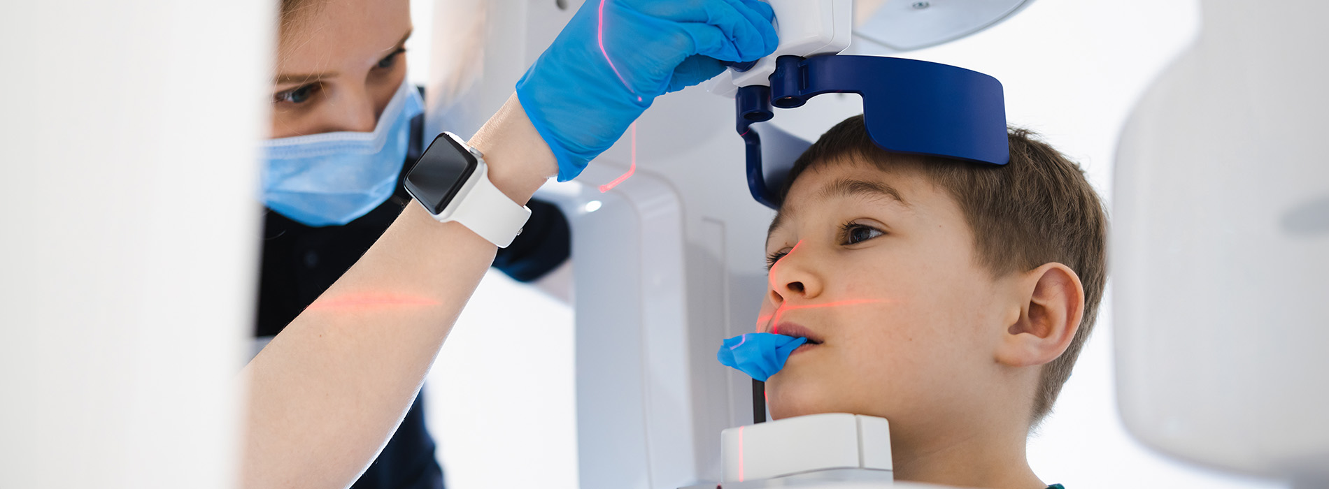
Digital X-rays use electronic sensors and computer processing to capture dental images instead of traditional film. The sensor—typically a small, flat electronic plate—is briefly placed in the mouth or positioned outside for panoramic views, and it converts X-ray energy into a digital file that appears on a monitor within seconds. That immediate feedback helps clinicians evaluate tooth development, detect hidden decay, and assess jaw growth more efficiently than film-based methods.
Because the image is a digital file, it can be enlarged, adjusted for contrast, and annotated without losing quality. These enhancements make small details easier to see and help dentists explain findings to parents and young patients. The ability to manipulate images on-screen also reduces the need for repeat exposures when a particular angle or detail must be reviewed.
At Amarillo Super Smiles For Kids we use digital imaging as a routine diagnostic tool because it streamlines care and supports better communication between our team, families, and any specialists involved in a child’s treatment plan. The technology is now a standard of care in modern pediatric dentistry for delivering accurate, timely assessments with minimal disruption to the visit.
One of the most important advantages of digital X-rays is the lower radiation dose compared with conventional film. Digital sensors are more sensitive to X-rays, which means clear diagnostic images can be produced with less radiation. In pediatric dentistry, where patients are more sensitive to ionizing radiation, this reduction is particularly valuable and aligned with the ALARA principle—keeping exposure As Low As Reasonably Achievable.
Clinicians tailor imaging frequency and type to each child’s needs, balancing diagnostic benefit against exposure. Rather than taking routine images at every visit, X-rays are used selectively based on age, risk of decay, symptoms, and developmental milestones. Lead aprons, thyroid collars, and modern machine shielding further limit exposure during the brief moment an image is captured.
Because digital imaging produces good-quality images with less radiation, it allows for safer monitoring of growth and development over time. Parents can be reassured that necessary radiographs are performed with careful consideration and contemporary safety measures in place.
Digital X-rays reveal problems that aren’t visible during a visual exam, such as interproximal cavities between teeth, early-stage tooth decay under existing fillings, and abnormalities in tooth eruption. For children whose dentition is changing rapidly, these images are essential for tracking tooth positions, identifying impacted teeth, and planning timely interventions that can prevent more extensive treatment later on.
Because images are available immediately, the dental team can discuss findings with families during the same appointment. Being able to show an enlarged, clear image of a concern helps caregivers understand the condition and the rationale for recommended care. This transparency supports informed decision-making and reduces uncertainty about next steps.
Digital radiography also streamlines coordination with specialists when needed. Files can be securely shared with orthodontists, oral surgeons, or other pediatric dental specialists to support collaborative treatment planning without sending physical film or waiting for mail—a benefit for care continuity and faster scheduling of follow-up procedures.
Most digital radiographic procedures are quick and straightforward. For intraoral images—bitewings or periapicals—the child may be asked to bite gently on a small sensor while the dental professional positions the X-ray head. Panoramic images, which capture the whole mouth in one shot, require the child to stand or sit still while a rotating arm moves around the head for a few seconds. The entire process usually takes only minutes.
Staff are trained to make the experience as comfortable and calm as possible for younger patients. Sensors come in sizes appropriate for infants, children, and adolescents, and positioning is adjusted to minimize discomfort. When necessary, behavior guidance techniques and a soothing environment help children relax so a clear image can be obtained without repeat exposures.
Once an image is captured it appears on a screen immediately. The dentist will review it, point out areas of interest, and explain what the image shows in straightforward language. If additional views are required for diagnosis, the team will take them promptly—digital systems make repeats fast while still maintaining careful attention to exposure minimization.
Digital images offer high resolution and the ability to enhance contrast, brightness, and zoom so clinicians can detect subtle signs of disease or developmental issues. These editing tools are purely for clinical interpretation and do not alter the original data; they simply make diagnostically relevant details easier to evaluate. Clear images help create precise treatment plans, monitor healing after procedures, and document long-term oral development.
Electronic records are stored in secure, HIPAA-compliant systems that protect patient privacy while allowing access by authorized providers involved in a child’s care. Secure sharing options facilitate timely consultations with specialists and make it easier to transfer records when families move or change providers, without the delay and environmental cost associated with physical films.
Beyond diagnosis, digital X-rays are a practical educational tool. The on-screen images help the dental team show parents and children exactly what is happening inside the mouth, explain preventive steps, and demonstrate the expected progress after treatment. That visual approach often improves understanding and supports better home care and follow-through.
In summary, digital X-rays are a fast, reliable, and safer way to obtain detailed dental images that support accurate diagnosis and thoughtful treatment planning for children. If you have questions about how digital radiography is used or what to expect during your child’s next visit, please contact us for more information and guidance.
Digital X-rays use electronic sensors and computer processing to capture images of teeth and jaws instead of traditional film. The images appear on a monitor within seconds, allowing dentists to evaluate tooth development, detect hidden decay, and assess jaw growth efficiently.
Digital X-rays produce images instantly, can be enlarged and adjusted for contrast, and require less radiation than traditional film. This allows for safer imaging, faster diagnosis, and easier sharing of images with specialists.
Yes. Digital X-rays use lower radiation doses compared with conventional film, and additional safety measures like lead aprons and thyroid collars are used. Dentists follow the ALARA principle, keeping exposure As Low As Reasonably Achievable.
They reveal problems not visible during a regular exam, such as cavities between teeth, decay under fillings, impacted teeth, or developmental abnormalities. These images help track growth and plan timely interventions to prevent more extensive treatments later.
For intraoral images, a small sensor is placed in the mouth while the X-ray head captures the image. Panoramic images require the child to sit or stand still while the machine rotates. The process is quick, usually only taking a few minutes, and is designed to be comfortable for children.
Digital images allow dentists to see fine details, plan precise treatments, monitor healing, and track long-term oral development. They can be enhanced on-screen for clarity without altering the original data, supporting accurate and informed decision-making.
Yes. Digital files can be securely shared with orthodontists, oral surgeons, or other pediatric dental specialists, facilitating collaborative treatment planning and faster follow-up care without sending physical film.
Not always. Dentists use X-rays selectively based on a child’s age, cavity risk, symptoms, and developmental milestones. This ensures imaging is performed only when necessary to support accurate diagnosis and treatment planning.
Yes. On-screen images help dentists explain dental conditions, demonstrate preventive steps, and show expected progress after treatment. This visual approach improves understanding and supports better home care and follow-through.
Digital images are stored securely in HIPAA-compliant systems, protecting patient privacy. Authorized providers can access the images as needed, and secure sharing options make it easy to consult specialists or transfer records if families move.
